Modelling cell-type proportions in repeated-measures designs with sccomp: a TMA case study

Mixed-effect models with random effects for tissue microarrays and other complex designs

Author
Affiliation

Stefano Mangiola

SAiGENCI, Adelaide University, Australia

Published

October 29, 2026

Contributor: Pierre-Paul Axisa, CBMR, Copenhagen, Denmark.

Introduction

Testing cell proportional differences from single-cell-resolved count data is central to modern tissue analyses. It becomes a repeated-measures problem as soon as several observations come from the same donor, such as multiple tumour cores in a tissue microarray (TMA) or serial biopsies over a time course. Averaging cores up to the patient and testing one phenotype at a time drops both the compositional constraint and the repeated-measures structure.

sccomp is a Bayesian model for mixed-effect compositional designs that identifies outliers probabilistically, tests for differential variability, and removes unwanted effects. It takes a long tibble with one row per sample and cell type, a count column, and the design columns, and sccomp_estimate(), sccomp_test(), and sccomp_predict() return tibbles ready for dplyr, tidyr, and ggplot2.

Source study and study design

We apply sccomp to the NeoTRIP trial. Wang et al. (2023) used imaging mass cytometry (IMC) to map how multicellular spatial organization in triple-negative breast cancer relates to neoadjuvant immunotherapy response. In the phase III NeoTRIP trial (NCT02620280), early high-risk TNBC patients were randomised 1:1 to carboplatin + nab-paclitaxel (C) or the same regimen plus atezolizumab (C&I) for eight 3-week neoadjuvant cycles before surgery. Research FFPE biopsies:

  • Baseline (n = 243)
  • On-treatment (first day of cycle 2; n = 207)
  • Post-treatment (surgical excision; n = 210)
  • Profiled with a 43-marker IMC panel at subcellular resolution
  • Deep-learning segmentation and semi-supervised clustering defined 17 epithelial and 20 TME cell phenotypes
  • Multiple regions of interest (ROIs) were imaged per biopsy

Each patient contributes several samples. The biopsies are nested in the patient, and the regions of interest are nested in the biopsy. Patient NT193, in the chemotherapy arm with residual disease, was imaged at all three timepoints: 10 ROIs at baseline, 10 on-treatment, and 3 at surgery. In the model, each ROI is one compositional sample and the patient is the repeated-measures group.

One patient, three timepoints, several ROIs
mk_spatial_tile <- function(dat, square = TRUE) {
  if (nrow(dat) == 0) {
    dat <- tibble::tibble(
      Location_Center_X = numeric(),
      Location_Center_Y = numeric(),
      draw = character()
    )
  }
  tile <- ggplot(dat, aes(x = Location_Center_X, y = Location_Center_Y)) +
    geom_point(data = filter(dat, draw == "other"), colour = "#39FF14", size = 0.10) +
    geom_point(data = filter(dat, draw == "pdl1"), colour = "#FFFF00", size = 0.16) +
    geom_point(data = filter(dat, draw == "m2"), colour = "#FF00FF", size = 0.16) +
    scale_x_continuous(labels = NULL, guide = "none", expand = c(0, 0)) +
    scale_y_reverse(labels = NULL, guide = "none", expand = c(0, 0)) +
    theme_void(base_size = 8) +
    theme(
      plot.background = element_rect(fill = "white", colour = NA),
      panel.background = element_rect(fill = "black", colour = NA),
      legend.position = "none",
      plot.margin = margin(
        if (square) 1 else 0, if (square) 1 else 0,
        if (square) 1 else 0, if (square) 1 else 0,
        unit = "pt"
      )
    )
  if (square) tile <- tile + theme(aspect.ratio = 1)
  tile
}

roi <- read_csv(
  file.path(results_dir, "sccomp_long_tme_immune_roi.csv"),
  col_select = c(PatientID, BiopsyPhase, ImageID),
  show_col_types = FALSE
) |>
  filter(PatientID == "NT193") |>
  distinct() |>
  mutate(BiopsyPhase = factor(BiopsyPhase, phase_levels)) |>
  arrange(BiopsyPhase, ImageID) |>
  mutate(i = row_number(), .by = BiopsyPhase)

n_phase <- roi |>
  summarise(n = n(), .by = BiopsyPhase) |>
  mutate(x0 = cumsum(n + 2) - (n + 2))
roi <- roi |>
  left_join(n_phase, by = "BiopsyPhase") |>
  mutate(x = i + x0)

cells_nt <- read_fst(file.path(post_dir, "nt193-cells.fst")) |>
  as_tibble()
dx <- 0.46
pad <- 0.62
xmin <- min(roi$x) - pad
xmax <- max(roi$x) + pad
fig_w <- 12
fig_h <- 5.4
target_in <- (2 * dx) / (xmax - xmin) * fig_w
headroom <- 1.9
tile_h <- target_in * headroom / (fig_h - target_in)

phase <- roi |>
  summarise(x = mean(x), .by = BiopsyPhase) |>
  mutate(
    y = tile_h + 0.62,
    label = case_match(
      as.character(BiopsyPhase),
      "Baseline" ~ "Baseline\n10 ROIs",
      "On-treatment" ~ "On-treatment\n10 ROIs",
      "Post-treatment" ~ "Post-treatment\n3 ROIs"
    )
  )
patient <- tibble::tibble(
  x = mean(phase$x), y = phase$y[1] + 0.95, label = "Donor"
)
edges <- bind_rows(
  phase |> mutate(x, y, xend = patient$x, yend = patient$y),
  roi |> mutate(
    x,
    y = tile_h,
    xend = phase$x[match(BiopsyPhase, phase$BiopsyPhase)],
    yend = phase$y[1]
  )
)
phase_cols <- c(
  Baseline = "#1B9E77",
  `On-treatment` = "#D95F02",
  `Post-treatment` = "#7570B3"
)

tree <- ggplot() +
  geom_segment(
    data = edges, aes(x = x, y = y, xend = xend, yend = yend),
    colour = "grey70", linewidth = 0.4
  ) +
  geom_label(
    data = phase,
    aes(x, y, label = label, fill = BiopsyPhase),
    colour = "white", fontface = "bold", size = 3, label.size = 0
  ) +
  geom_label(
    data = patient, aes(x, y, label = label),
    fill = "grey15", colour = "white", fontface = "bold", size = 3.6, label.size = 0
  ) +
  scale_fill_manual(values = phase_cols, guide = "none") +
  coord_cartesian(
    xlim = c(xmin, xmax),
    ylim = c(-0.04, patient$y + 0.4),
    clip = "off"
  ) +
  labs(title = "Patient NT193") +
  theme_void(base_size = 11) +
  theme(
    plot.title = element_text(face = "bold", hjust = 0.5, size = 12),
    plot.margin = margin(6, 6, 6, 6)
  )

for (i in seq_len(nrow(roi))) {
  tree <- tree + annotation_custom(
    ggplotGrob(mk_spatial_tile(filter(cells_nt, ImageID == roi$ImageID[i]), square = FALSE)),
    xmin = roi$x[i] - dx,
    xmax = roi$x[i] + dx,
    ymin = 0,
    ymax = tile_h
  )
}
tree
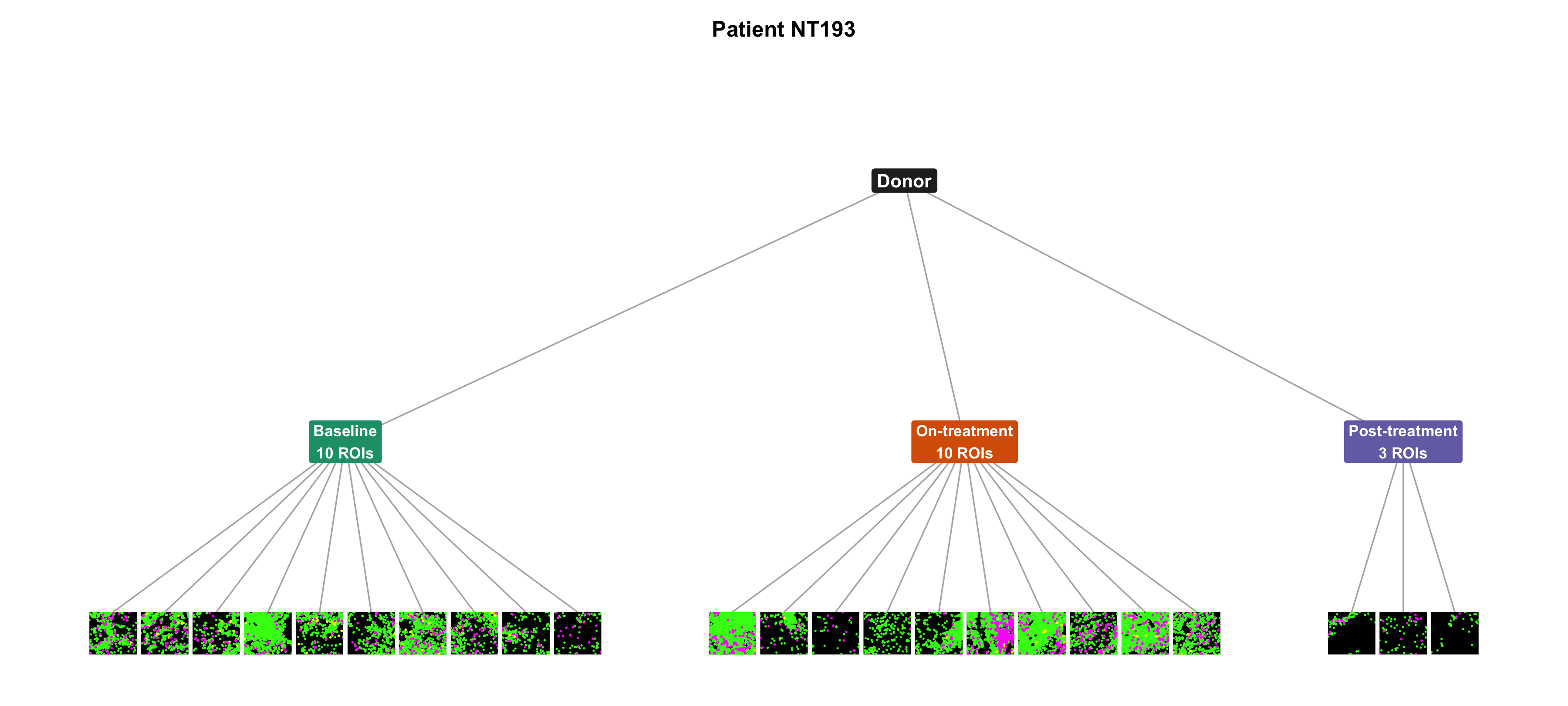
Tree for patient NT193. The patient sits at the top and three biopsy timepoints branch underneath. Each timepoint ends in the imaged regions of interest, drawn as spatial tiles: magenta M2 macrophages, yellow PD-L1-positive antigen-presenting cells, and green other immune cells. There are 10 tiles at baseline, 10 on-treatment, and 3 post-treatment.
Figure 1

Wang et al. linked cell densities, spatial interactions, and proliferative fractions to pathological complete response (pCR), and showed that immunotherapy remodels tissue composition over time, with response-specific dynamics of immune infiltration (notably in their Fig. 5 compositional trajectory analysis).

Quantity Value
Per-protocol patients 257
ROI-level immune samples 1,740
Immune cell phenotypes 15
Median ROIs per patient 7

sccomp mixed-effects compositional modelling

Wang et al. summarised longitudinal composition as mean phenotype proportions per patient and compared arms, timepoints, and response groups with Wilcoxon tests, Z-scored trend plots (their Fig. 5), and per-phenotype weighted binomial GLMs on tumour-level proportions. Those tests treat each phenotype in isolation and average ROIs to the patient before inference, setting aside compositional closure (proportions sum to one) and the nesting of biopsies and ROIs within patients.

With sccomp, we model the ROI-level cell counts directly on a latent log-ratio scale with mixed effects on the mean and variability. Each ROI is one compositional sample (sample = "ROI_id" in sccomp_estimate()), several ROIs share a biopsy timepoint (BiopsyPhase), and a patient random intercept (1 | PatientID) accounts for baseline differences across individuals and shrinkage across the cohort:

\[\text{composition} \sim \text{Arm} \times \text{BiopsyPhase} \times \text{pCR} + (1 \mid \text{PatientID})\]

sccomp analysis

The blocks below are the minimal tidyverse code to reproduce the analysis locally from the parsed input (run top to bottom), using the immune TME compartment with ROI-level samples and a patient random intercept.

From long-format tidy input

The input long is a tibble with one row per ROI × immune phenotype: ROI_id, a count, the design columns (Arm, BiopsyPhase, pCR), and the grouping PatientID. Several ROIs share one BiopsyPhase. Preview:

Load long-format input
library(readr)
library(dplyr)

long <- read_csv(file.path(results_dir, "sccomp_long_tme_immune_roi.csv"),
                 show_col_types = FALSE) |>
  rename(ROI_id = sample) |>
  mutate(
    count = as.integer(count),
    BiopsyPhase = factor(BiopsyPhase, c("Baseline", "On-treatment", "Post-treatment")),
    Arm = factor(Arm, c("C", "C&I")),
    pCR = factor(pCR, c("RD", "pCR"))
  )

long |>
  select(ROI_id, cell_group, count, PatientID, BiopsyPhase, Arm, pCR) |>
  gt::gt_preview() |>
  gt::tab_caption("Long-format sccomp input (1,740 ROIs x 15 immune phenotypes)")
Long-format sccomp input (1,740 ROIs x 15 immune phenotypes)
ROI_id cell_group count PatientID BiopsyPhase Arm pCR
1 NTImg0011 CD8^+T 19 NT031 Baseline C&I RD
2 NTImg0011 CD8^+GZMB^+T 1 NT031 Baseline C&I RD
3 NTImg0011 CD8^+TCF1^+T 1 NT031 Baseline C&I RD
4 NTImg0011 CD8^+PD1^+T_{Ex} 7 NT031 Baseline C&I RD
5 NTImg0011 CD4^+TCF1^+T 4 NT031 Baseline C&I RD
6..26099
26100 NTImg1889 PD-L1^+IDO^+APCs 71 NT091 On-treatment C&I RD

Fit the model

library(sccomp)

fit <- sccomp_estimate(
  long,
  formula_composition = ~ Arm * BiopsyPhase * pCR + (1 | PatientID),
  formula_variability = ~ 1,
  sample = "ROI_id",
  cell_group = "cell_group",
  abundance = "count",
  inference_method = "hmc",
  cores = parallel::detectCores(),
  verbose = FALSE
) |>
  sccomp_test()
Fit and test the model
library(sccomp)

fit <- sccomp_estimate(
  long,
  formula_composition = ~ Arm * BiopsyPhase * pCR + (1 | PatientID),
  formula_variability = ~ 1,
  sample = "ROI_id",
  cell_group = "cell_group",
  abundance = "count",
  inference_method = "hmc",
  cores = parallel::detectCores(),
  verbose = FALSE
) |>
  sccomp_test()
sccomp model
============

Model specifications:
  Family: multi_beta_binomial 
  Composition formula: ~Arm * BiopsyPhase * pCR + (1 | PatientID) 
  Variability formula: ~1 
  Inference method: hmc 

Data: Samples: 1740   Cell groups: 15 

Column prefixes: c_ -> composition parameters  v_ -> variability parameters

Convergence diagnostics:
  For each parameter, n_eff is the effective sample size and R_k_hat is the potential
  scale reduction factor on split chains (at convergence, R_k_hat = 1).

# A tibble: 4,035 × 19
   cell_group parameter  factor c_lower c_effect  c_upper   c_pH0   c_FDR c_rhat
   <chr>      <chr>      <chr>    <dbl>    <dbl>    <dbl>   <dbl>   <dbl>  <dbl>
 1 CD20^+B    (Intercep… <NA>   -0.710   -0.514  -0.314   0       0         1.00
 2 CD20^+B    ArmC&I     Arm    -0.579   -0.292  -0.00208 0.0911  3.66e-2   1.00
 3 CD20^+B    BiopsyPha… Biops… -0.0970   0.134   0.359   0.377   1.30e-1   1.00
 4 CD20^+B    BiopsyPha… Biops…  0.903    1.12    1.33    0       0         1.00
 5 CD20^+B    pCRpCR     pCR    -0.162    0.112   0.390   0.465   2.21e-1   1.00
 6 CD20^+B    ArmC&I:Bi… <NA>   -0.281    0.0545  0.396   0.602   3.17e-1   1.00
 7 CD20^+B    ArmC&I:Bi… <NA>   -0.479   -0.151   0.189   0.384   1.24e-1   1.00
 8 CD20^+B    ArmC&I:pC… <NA>   -0.227    0.164   0.540   0.368   2.55e-1   1.00
 9 CD20^+B    BiopsyPha… <NA>   -0.274    0.0505  0.377   0.625   3.86e-1   1.00
10 CD20^+B    BiopsyPha… <NA>   -0.897   -0.572  -0.259   0.00175 4.17e-4   1.00
# ℹ 4,025 more rows
# ℹ 10 more variables: c_ess_bulk <dbl>, c_ess_tail <dbl>, v_lower <dbl>,
#   v_effect <dbl>, v_upper <dbl>, v_pH0 <dbl>, v_FDR <dbl>, v_rhat <dbl>,
#   v_ess_bulk <dbl>, v_ess_tail <dbl>

sccomp_proportional_fold_change() reports how fitted proportions change between two conditions. The result is descriptive; significance still comes from sccomp_test(). Here, chemoimmunotherapy at surgery, residual disease versus pathological complete response:

Proportional fold change
fit |>
  sccomp_proportional_fold_change(
    formula_composition = ~ Arm * BiopsyPhase * pCR,
    from = "C&I:Post-treatment:RD",  # chemoimmunotherapy, surgery, residual disease
    to = "C&I:Post-treatment:pCR"    # same arm and visit, pathological complete response
  ) |>
  gt::gt_preview() |>
  gt::tab_caption(
    "Proportional fold change: C&I post-treatment, pCR versus residual disease"
  )
Running standalone generated quantities after 1 MCMC chain, with 1 thread(s) per chain...

Chain 1  Elapsed Time: 0.1 seconds (Generated Quantities) 
Chain 1 finished in 0.0 seconds.
Proportional fold change: C&I post-treatment, pCR versus residual disease
cell_group proportion_fold_change average_uncertainty statement
1 CD20^+B -1.300258 0.02649652 1.3-fold decrease (from 0.0645 to 0.0496)
2 CD4^+PD1^+T -1.595534 0.04749542 1.6-fold decrease (from 0.0439 to 0.0275)
3 CD4^+TCF1^+T -1.100683 0.01633892 1.1-fold decrease (from 0.0918 to 0.0834)
4 CD56^+NK 1.571215 0.04918382 1.6-fold increase (from 0.0808 to 0.1269)
5 CD79a^+Plasma 1.132715 0.02740352 1.1-fold increase (from 0.0721 to 0.0816)
6..14
15 Treg -3.406119 0.25870011 3.4-fold decrease (from 0.0275 to 0.0081)

Model checks

Convergence. All 4,035 composition parameters have median \(\hat{R}\) = 1.001 (maximum 1.014). Bulk effective sample size has median 4,837 (minimum 495). Tail effective sample size has median 2,850 (minimum 923).

Variability is an intercept for each phenotype. Those 15 coefficients have maximum \(\hat{R}\) = 1.003, minimum bulk ESS 3,404, and minimum tail ESS 2,451. The composition parameters with the smallest bulk ESS:

Smallest bulk effective sample sizes
fit |>
  filter(is.finite(c_ess_bulk)) |>
  slice_min(c_ess_bulk, n = 6, with_ties = FALSE) |>
  gt::gt_preview() |>
  gt::tab_caption(
    "Composition parameters with the smallest bulk effective sample size"
  )
Composition parameters with the smallest bulk effective sample size
cell_group parameter factor c_lower c_effect c_upper c_pH0 c_FDR c_rhat c_ess_bulk c_ess_tail v_lower v_effect v_upper v_pH0 v_FDR v_rhat v_ess_bulk v_ess_tail
1 DCs ArmC&I Arm -0.08375482 0.1601228 0.399447699 0.31431431 0.148577149 1.009882 600.7795 1411.607 NA NA NA NA NA NA NA NA
2 DCs (Intercept) NA 0.77417609 0.9424418 1.116479608 0.00000000 0.000000000 1.011012 697.7882 1552.972 -3.78283 -2.957539 -2.134048 0 0 1.001649 4598.984 2582.758
3 DCs ArmC&I:pCRpCR NA -0.47084651 -0.1203059 0.229162709 0.45070070 0.295827077 1.010088 717.1771 1444.238 NA NA NA NA NA NA NA NA
4 M2 Mac ArmC&I Arm 0.12079694 0.3597968 0.601198700 0.01701702 0.009384384 1.019933 776.9721 1483.882 NA NA NA NA NA NA NA NA
5 DCs pCRpCR pCR -0.52733358 -0.2649256 -0.008725984 0.10010010 0.057807808 1.007276 858.1370 1509.393 NA NA NA NA NA NA NA NA
6 M2 Mac ArmC&I:pCRpCR NA -0.74712725 -0.4227944 -0.071407724 0.03678679 0.036786787 1.008612 878.2435 1583.149 NA NA NA NA NA NA NA NA

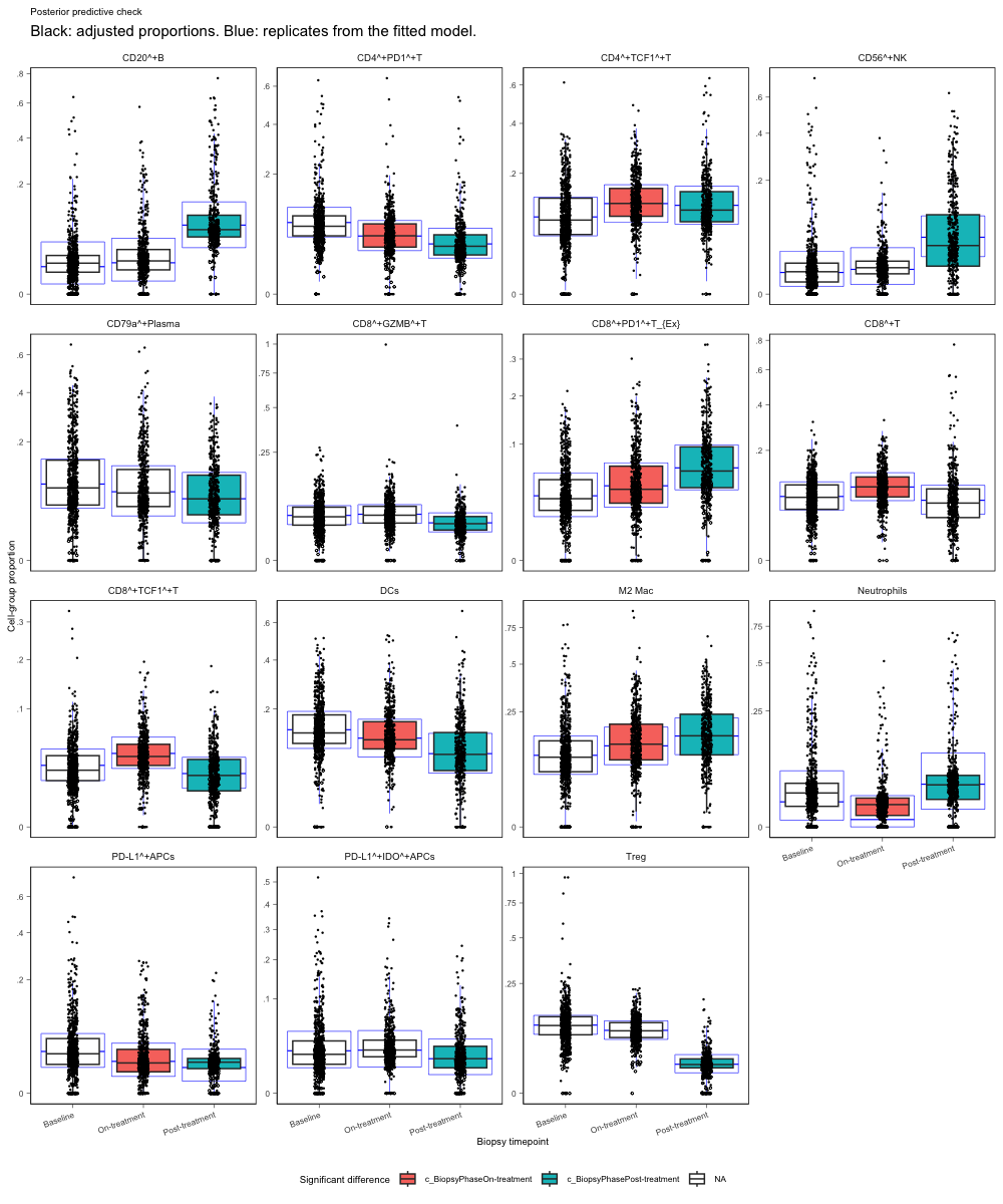
Posterior predictive check. remove_unwanted_effects = TRUE removes variation other than biopsy timepoint, so the black boxes are adjusted proportions and the blue boxes are posterior predictive replicates. Fill marks a timepoint contrast with FDR below 0.05.

fit |> 
  sccomp_boxplot(
  factor = "BiopsyPhase",
  significance_statistic = "FDR",
  remove_unwanted_effects = TRUE
  ) +
  labs(
    title = "Posterior predictive check",
    subtitle = "Black: adjusted proportions. Blue: replicates from the fitted model.",
    x = "Biopsy timepoint"
  ) +
  guides(colour = "none", shape = "none") 

Boxplots of immune-cell proportions by biopsy timepoint, faceted by cell type, after removing variation other than biopsy timepoint. Black boxes are adjusted proportions and blue boxes are posterior predictive replicates. Filled boxes mark a timepoint contrast with FDR below 0.05.

Hypothesis testing

We pass contrasts to sccomp_test() as strings.

marginal_CI_post compares responders with residual disease at that visit alone. Any gap the two groups already had at baseline is still in this result.

did_CI_post compares how far each group moved from its own baseline. A head start cancels, so the contrast is the difference in change. We use this contrast because a visit-only comparison still carries the baseline gap. Post-treatment M2 proportions are similar in the two response groups, so that snapshot does not describe the change.

Test two contrasts
contrasts <- c(
  marginal_CI_post = paste(
    "`pCRpCR`",                                  # pCR versus RD in chemotherapy at baseline
    "`ArmC&I:pCRpCR`",                           # additional pCR versus RD gap in the C&I arm
    "`BiopsyPhasePost-treatment:pCRpCR`",        # additional pCR versus RD gap at post-treatment
    "`ArmC&I:BiopsyPhasePost-treatment:pCRpCR`", # additional pCR versus RD gap for C&I at post-treatment
    sep = " + "
  ),
  did_CI_post = paste(
    "`BiopsyPhasePost-treatment:pCRpCR`",        # change from baseline in the pCR contrast
    "`ArmC&I:BiopsyPhasePost-treatment:pCRpCR`", # further change from baseline in the C&I arm
    sep = " + "
  )
)

res <- sccomp_test(fit, contrasts = contrasts)

res holds those two contrasts, one row per phenotype. The table is the full set of difference-in-differences, strongest first:

Preview difference-in-differences contrasts
hmc_did |>
  filter(contrast_family == "did") |>
  arrange(c_FDR) |>
  gt::gt_preview() |>
  gt::tab_caption("Change from baseline (strongest by FDR)")
Change from baseline (strongest by FDR)
parameter cell_group factor c_lower c_effect c_upper c_pH0 c_FDR c_rhat c_ess_bulk c_ess_tail contrast_family Arm BiopsyPhase pCR
1 did__C&I__Post_treatment CD56^+NK NA 2.4152900 3.25669670 4.1111366 0.0000000 0.0000000 1.0010947 4247.316 3275.396 did C&I Post-treatment pCR_minus_RD
2 did__C&I__Post_treatment DCs NA 0.6045030 1.33237959 2.0188092 0.0000000 0.0000000 0.9996942 5122.662 3061.238 did C&I Post-treatment pCR_minus_RD
3 did__C&I__Post_treatment Neutrophils NA 1.6664088 2.63412173 3.5857767 0.0000000 0.0000000 1.0017040 4446.958 3336.609 did C&I Post-treatment pCR_minus_RD
4 did__C&I__Post_treatment PD-L1^+APCs NA -3.9597930 -2.80065090 -1.6193615 0.0000000 0.0000000 1.0002327 3677.553 2867.557 did C&I Post-treatment pCR_minus_RD
5 did__C&I__Post_treatment Treg NA -2.8820452 -1.94723055 -1.0560065 0.0000000 0.0000000 1.0006019 2449.355 2366.402 did C&I Post-treatment pCR_minus_RD
6..59
60 did__C&I__On_treatment CD8^+TCF1^+T NA -0.3600534 0.01963656 0.4067838 0.6599099 0.2803971 1.0012927 5303.619 3036.666 did C&I On-treatment pCR_minus_RD

Population and per-patient predictions

Population prediction grid
# One row per arm, timepoint, and response group.
pop <- tibble::tribble(
  ~Arm,  ~BiopsyPhase,     ~pCR,
  "C",   "Baseline",       "RD",
  "C",   "Baseline",       "pCR",
  "C",   "On-treatment",   "RD",
  "C",   "On-treatment",   "pCR",
  "C",   "Post-treatment", "RD",
  "C",   "Post-treatment", "pCR",
  "C&I", "Baseline",       "RD",
  "C&I", "Baseline",       "pCR",
  "C&I", "On-treatment",   "RD",
  "C&I", "On-treatment",   "pCR",
  "C&I", "Post-treatment", "RD",
  "C&I", "Post-treatment", "pCR"
) |>
  mutate(
    Arm = factor(Arm, c("C", "C&I")),
    BiopsyPhase = factor(BiopsyPhase, c("Baseline", "On-treatment", "Post-treatment")),
    pCR = factor(pCR, c("RD", "pCR")),
    ROI_id = paste0("pred_", row_number()),
    PatientID = "NT031"
  )

gt::gt_preview(pop) |>
  gt::tab_caption("Population grid passed to sccomp_predict()")
Population grid passed to sccomp_predict()
Arm BiopsyPhase pCR ROI_id PatientID
1 C Baseline RD pred_1 NT031
2 C Baseline pCR pred_2 NT031
3 C On-treatment RD pred_3 NT031
4 C On-treatment pCR pred_4 NT031
5 C Post-treatment RD pred_5 NT031
6..11
12 C&I Post-treatment pCR pred_12 NT031
Predict population proportions
pred_pop <- sccomp_predict(fit, new_data = pop)
Running standalone generated quantities after 1 MCMC chain, with 1 thread(s) per chain...

Chain 1  Elapsed Time: 0.185 seconds (Generated Quantities) 
Chain 1 finished in 0.0 seconds.
Predict population proportions
pred_pop |>
  gt::gt_preview() |>
  gt::tab_caption("Population predicted proportions")
Population predicted proportions
Arm BiopsyPhase pCR ROI_id PatientID cell_group proportion_mean proportion_lower proportion_upper unconstrained_mean unconstrained_lower unconstrained_upper
1 C Baseline RD pred_1 NT031 CD20^+B 0.025475434 0.011849041 0.04656918 -0.78916098 -1.44397950 -0.18577897
2 C Baseline RD pred_1 NT031 CD4^+PD1^+T 0.050589704 0.032262354 0.07256136 -0.07387369 -0.48652249 0.30169921
3 C Baseline RD pred_1 NT031 CD4^+TCF1^+T 0.081006649 0.051677388 0.11175873 0.39988733 -0.02541777 0.76499292
4 C Baseline RD pred_1 NT031 CD56^+NK 0.032822597 0.017883680 0.05537661 -0.52534173 -1.06513952 0.01087662
5 C Baseline RD pred_1 NT031 CD79a^+Plasma 0.158685350 0.102258774 0.22280513 1.07227269 0.63309480 1.48362000
6..179
180 C&I Post-treatment pCR pred_12 NT031 Treg 0.008723248 0.005836047 0.01223340 -1.53308238 -1.88980277 -1.19372516
Per-patient prediction grid
# Per-patient grid: one row per patient and timepoint (uses the patient random intercept)
pat <- long |>
  distinct(PatientID, BiopsyPhase, Arm, pCR) |>
  mutate(ROI_id = paste0(PatientID, "_", BiopsyPhase))

pat |>
  gt::gt_preview() |>
  gt::tab_caption("Per-patient prediction grid (624 rows)")
Per-patient prediction grid (624 rows)
PatientID BiopsyPhase Arm pCR ROI_id
1 NT031 Baseline C&I RD NT031_Baseline
2 NT277 Baseline C&I RD NT277_Baseline
3 NT277 On-treatment C&I RD NT277_On-treatment
4 NT238 Baseline C&I RD NT238_Baseline
5 NT238 On-treatment C&I RD NT238_On-treatment
6..623
624 NT091 On-treatment C&I RD NT091_On-treatment
Predict per-patient proportions
pred_pat <- sccomp_predict(fit, new_data = pat)
Running standalone generated quantities after 1 MCMC chain, with 1 thread(s) per chain...

Chain 1  Elapsed Time: 5.094 seconds (Generated Quantities) 
Chain 1 finished in 0.0 seconds.
Predict per-patient proportions
pred_pat
# A tibble: 9,360 × 12
   PatientID BiopsyPhase Arm   pCR   ROI_id         cell_group   proportion_mean
   <chr>     <fct>       <fct> <fct> <chr>          <chr>                  <dbl>
 1 NT031     Baseline    C&I   RD    NT031_Baseline CD20^+B               0.0175
 2 NT031     Baseline    C&I   RD    NT031_Baseline CD4^+PD1^+T           0.0427
 3 NT031     Baseline    C&I   RD    NT031_Baseline CD4^+TCF1^+T          0.0848
 4 NT031     Baseline    C&I   RD    NT031_Baseline CD56^+NK              0.0353
 5 NT031     Baseline    C&I   RD    NT031_Baseline CD79a^+Plas…          0.139 
 6 NT031     Baseline    C&I   RD    NT031_Baseline CD8^+GZMB^+T          0.0272
 7 NT031     Baseline    C&I   RD    NT031_Baseline CD8^+PD1^+T…          0.0420
 8 NT031     Baseline    C&I   RD    NT031_Baseline CD8^+T                0.0972
 9 NT031     Baseline    C&I   RD    NT031_Baseline CD8^+TCF1^+T          0.0477
10 NT031     Baseline    C&I   RD    NT031_Baseline DCs                   0.123 
# ℹ 9,350 more rows
# ℹ 5 more variables: proportion_lower <dbl>, proportion_upper <dbl>,
#   unconstrained_mean <dbl>, unconstrained_lower <dbl>,
#   unconstrained_upper <dbl>

Results

Predicted immune compositional trajectories

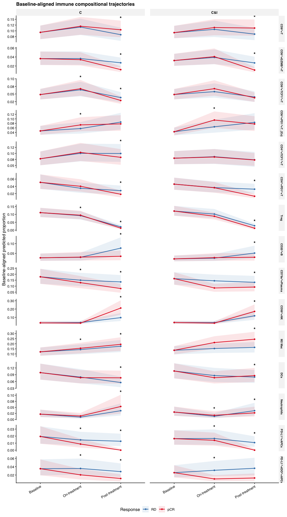
Figure 1 shows baseline-aligned predicted proportions (95% credible intervals) for all 15 immune TME phenotypes. Within each arm × cell-type facet, both response groups are shifted to the mean of their baseline predictions. Panels are split by treatment arm (columns); colour denotes pathological response. Asterisks mark arm–time points with difference-in-differences FDR < 0.05.

Align groups to a common baseline
# Plot shift. Within each arm and cell type, move pCR and RD to the mean of
# their baseline predictions, on the posterior draws.
draws <- sccomp_predict(
  fit, new_data = pop, number_of_draws = 500, summary_instead_of_draws = FALSE
) |>
  as_tibble() |>
  mutate(across(c(Arm, BiopsyPhase, pCR), as.character))
Running standalone generated quantities after 1 MCMC chain, with 1 thread(s) per chain...

Chain 1  Elapsed Time: 0.183 seconds (Generated Quantities) 
Chain 1 finished in 0.0 seconds.
Align groups to a common baseline
# Shift pCR and RD to their average baseline within each Arm x cell_group, per draw
shifts <- draws |>
  filter(BiopsyPhase == "Baseline") |>
  select(Arm, cell_group, .draw, pCR, proportion) |>
  pivot_wider(names_from = pCR, values_from = proportion) |>
  mutate(shift_pCR = (pCR + RD) / 2 - pCR,
         shift_RD  = (pCR + RD) / 2 - RD) |>
  select(Arm, cell_group, .draw, shift_pCR, shift_RD)

aligned_summary <- draws |>
  left_join(shifts, by = c("Arm", "cell_group", ".draw")) |>
  mutate(aligned_prop = proportion + if_else(pCR == "pCR", shift_pCR, shift_RD)) |>
  summarise(
    aligned_mean  = mean(aligned_prop),
    aligned_lower = quantile(aligned_prop, 0.025),
    aligned_upper = quantile(aligned_prop, 0.975),
    .by = c(Arm, BiopsyPhase, pCR, cell_group)
  )
Baseline-aligned immune trajectories
cell_order <- c(
  "CD8^+T", "CD8^+GZMB^+T", "CD8^+TCF1^+T", "CD8^+PD1^+T_{Ex}",
  "CD4^+TCF1^+T", "CD4^+PD1^+T", "Treg", "CD20^+B", "CD79a^+Plasma",
  "CD56^+NK", "M2 Mac", "DCs", "Neutrophils", "PD-L1^+APCs", "PD-L1^+IDO^+APCs"
)
cols_response <- c(RD = "#377EB8", pCR = "#E41A1C")

pred_main <- aligned_summary |>
  mutate(
    BiopsyPhase = factor(BiopsyPhase, phase_levels),
    Arm = factor(Arm, arm_levels),
    pCR = factor(pCR, c("RD", "pCR")),
    cell_group = factor(cell_group, levels = cell_order),
    # A few intervals sit just below zero after the baseline shift.
    aligned_mean = pmax(aligned_mean, 0),
    aligned_lower = pmax(aligned_lower, 0)
  )

# Asterisk just above the ribbon where the DiD FDR is below 0.05.
did_sig_fig1 <- hmc_did |>
  filter(contrast_family == "did", is.finite(c_FDR), c_FDR < 0.05) |>
  distinct(cell_group, Arm, BiopsyPhase) |>
  mutate(cell_group = factor(cell_group, levels = cell_order)) |>
  inner_join(
    pred_main |>
      summarise(ymax = max(aligned_upper), .by = c(cell_group, Arm, BiopsyPhase)),
    by = c("cell_group", "Arm", "BiopsyPhase")
  )

ggplot(pred_main, aes(x = BiopsyPhase, y = aligned_mean, colour = pCR, group = pCR)) +
  geom_ribbon(
    aes(ymin = aligned_lower, ymax = aligned_upper, fill = pCR),
    alpha = 0.10, colour = NA
  ) +
  geom_line(linewidth = 0.75) +
  geom_point(size = 1.2) +
  geom_text(
    data = did_sig_fig1,
    aes(x = BiopsyPhase, y = ymax, label = "*"),
    inherit.aes = FALSE,
    size = 4.5, colour = "black", fontface = "bold", vjust = -0.2
  ) +
  facet_grid(cell_group ~ Arm, scales = "free_y") +
  scale_y_continuous(labels = scales::label_number(accuracy = 0.01)) +
  coord_cartesian(clip = "off") +
  scale_colour_manual(values = cols_response, name = "Response") +
  scale_fill_manual(values = cols_response, name = "Response") +
  labs(
    title = "Baseline-aligned immune compositional trajectories",
    x = NULL,
    y = "Baseline-aligned predicted proportion"
  ) +
  theme_pub() +
  theme(strip.text.y = element_text(size = 7))
Line plots of baseline-aligned immune proportions across biopsy timepoints, faceted by cell type and treatment arm, with pathological complete response and residual disease in separate colours and asterisks where the difference-in-differences FDR is below 0.05.
Figure 2

Change from baseline

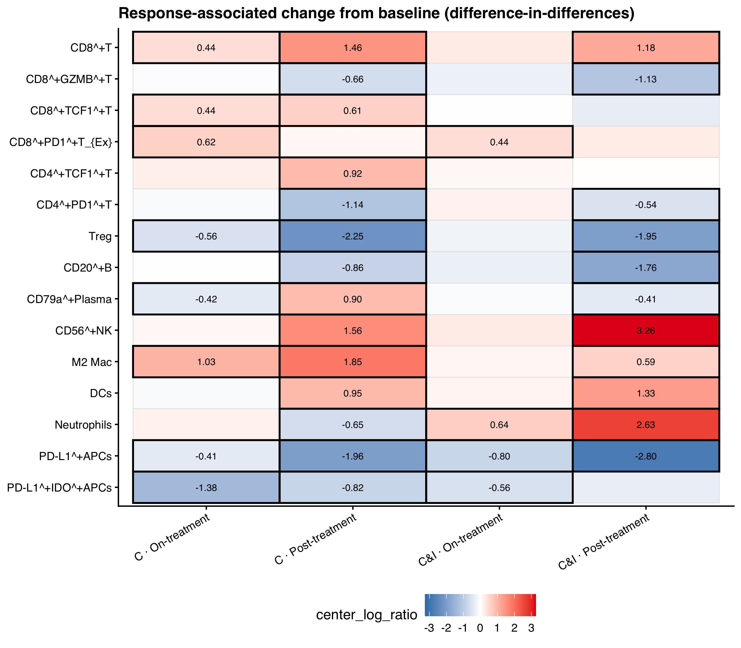
For each cell type and treatment arm, we test whether responders and patients with residual disease changed by different amounts from baseline. Figure 2 shows that difference at the on-treatment and post-treatment visits. Bordered tiles have FDR < 0.05.

Difference-in-differences heatmap
did_hm <- hmc_did |>
  filter(contrast_family == "did") |>
  mutate(
    cell_group = factor(cell_group, levels = rev(cell_order)),
    BiopsyPhase = factor(BiopsyPhase, levels = c("On-treatment", "Post-treatment")),
    Arm = factor(Arm, arm_levels)
  ) |>
  filter(!is.na(cell_group)) |>
  mutate(
    sig = is.finite(c_FDR) & c_FDR < 0.05,
    facet_lab = factor(
      paste(Arm, BiopsyPhase, sep = " · "),
      levels = c(
        "C · On-treatment", "C · Post-treatment",
        "C&I · On-treatment", "C&I · Post-treatment"
      )
    )
  )

lim <- max(abs(did_hm$c_effect), na.rm = TRUE)

ggplot(did_hm, aes(x = facet_lab, y = cell_group, fill = c_effect)) +
  geom_tile(colour = "grey90", linewidth = 0.3) +
  geom_tile(
    data = filter(did_hm, sig),
    colour = "black", linewidth = 0.7, fill = NA
  ) +
  geom_text(
    aes(label = if_else(sig, sprintf("%.2f", c_effect), "")),
    size = 2.6
  ) +
  scale_fill_gradient2(
    low = cols_response[["RD"]], mid = "white", high = cols_response[["pCR"]],
    midpoint = 0, limits = c(-lim, lim), name = "center_log_ratio"
  ) +
  labs(
    title = "Response-associated change from baseline (difference-in-differences)",
    x = NULL, y = NULL
  ) +
  theme_pub() +
  theme(
    axis.text.x = element_text(angle = 30, hjust = 1),
    panel.grid = element_blank()
  )
Heatmap of difference-in-differences effects, on the centered log-ratio scale, for each immune phenotype, treatment arm, and post-baseline visit. Black borders mark FDR below 0.05.
Figure 3

Finding: Under the difference-in-differences contrast, 14 immune phenotypes change from baseline at post-treatment, and 9 at the on-treatment visit.

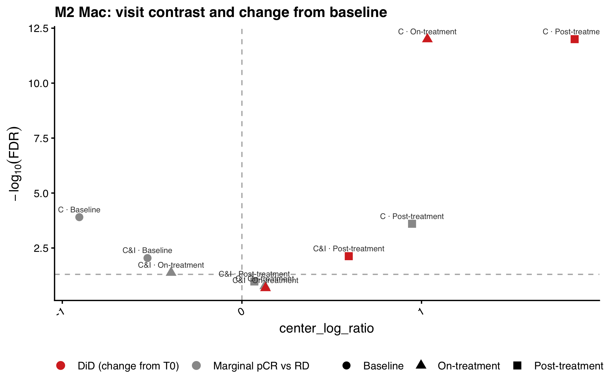
Finding: In the chemoimmunotherapy arm at post-treatment, M2 macrophages increase more from baseline in patients who reach pathological complete response (FDR 0.007), while PD-L1-positive antigen-presenting cells decrease (FDR < 0.0002).

Finding: The M2 macrophage change is not detected at the on-treatment visit (FDR 0.21).

M2 macrophage changes by response

Figure 3 compares pCR versus residual disease at each visit with the change from baseline. Points above the dotted line are FDR < 0.05.

M2 macrophage changes by response
m2_volcano <- bind_rows(
  hmc_marginal_pcr |>
    filter(cell_group == "M2 Mac") |>
    mutate(Arm, BiopsyPhase, c_effect, c_FDR, family = "Marginal pCR vs RD"),
  hmc_did |>
    filter(contrast_family == "did", cell_group == "M2 Mac") |>
    mutate(Arm, BiopsyPhase, c_effect, c_FDR, family = "DiD (change from T0)")
) |>
  mutate(label = paste(Arm, BiopsyPhase, sep = " · "))

ggplot(m2_volcano, aes(x = c_effect, y = -log10(pmax(c_FDR, 1e-12)))) +
  geom_vline(xintercept = 0, linetype = 2, colour = "grey70") +
  geom_hline(yintercept = -log10(0.05), linetype = 2, colour = "grey70") +
  geom_point(aes(colour = family, shape = BiopsyPhase), size = 2.8) +
  geom_text(aes(label = label), size = 2.3, vjust = -0.9, colour = "grey25") +
  scale_colour_manual(values = c(
    "Marginal pCR vs RD" = "#999999",
    "DiD (change from T0)" = "#d73027"
  )) +
  labs(
    title = "M2 Mac: visit contrast and change from baseline",
    x = "center_log_ratio",
    y = expression(-log[10](FDR)),
    colour = NULL, shape = NULL
  ) +
  theme_pub()
Scatter plot of M2 macrophage response contrasts, with the centered log-ratio effect on the x-axis and minus log10 FDR on the y-axis, comparing pCR versus residual disease at each visit with the change from baseline.
Figure 4

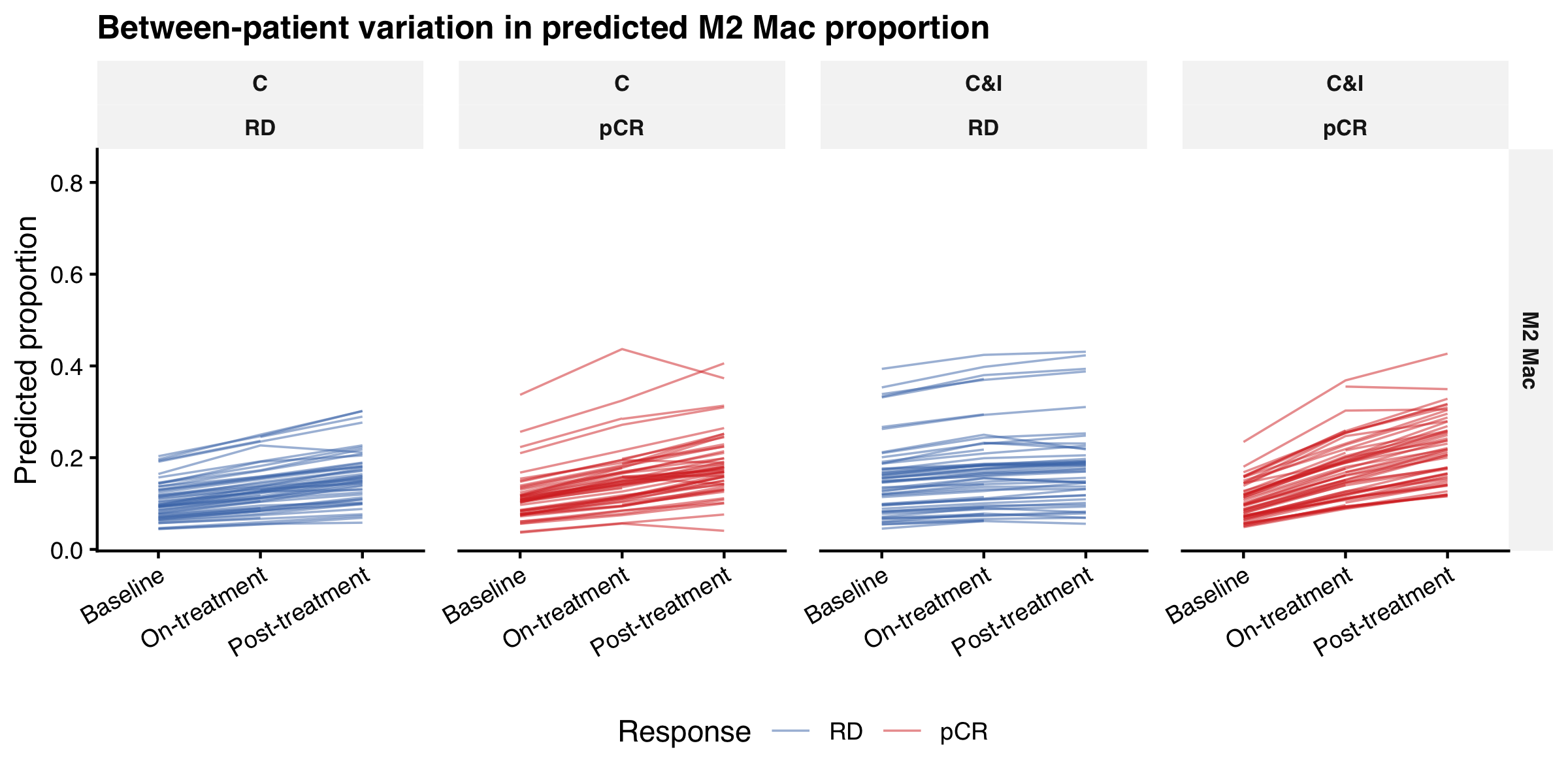
Between-patient heterogeneity in M2 Mac

The fixed effect is the population estimate. Patient random intercepts describe individual baselines and trajectories. Figure 4 shows the per-patient predicted M2 Mac trajectories.

Between-patient M2 trajectories
pred_pat <- hmc_pred_patient |>
  filter(cell_group == "M2 Mac") |>
  mutate(cell_group = factor(cell_group))

pcr_cols <- c("RD" = "#4575b4", "pCR" = "#d73027")

ggplot(pred_pat, aes(x = BiopsyPhase, y = proportion_mean, group = PatientID, colour = pCR)) +
  geom_line(linewidth = 0.4, alpha = 0.5) +
  facet_grid(cell_group ~ Arm + pCR, scales = "free_y") +
  scale_colour_manual(values = pcr_cols, name = "Response") +
  labs(
    title = "Between-patient variation in predicted M2 Mac proportion",
    x = NULL,
    y = "Predicted proportion"
  ) +
  theme_pub() +
  theme(legend.position = "bottom", strip.text = element_text(size = 8))
Line plots of predicted M2 macrophage proportion for each patient across biopsy timepoints, faceted by treatment arm and pathological response.
Figure 5

Visualise results in spatial context

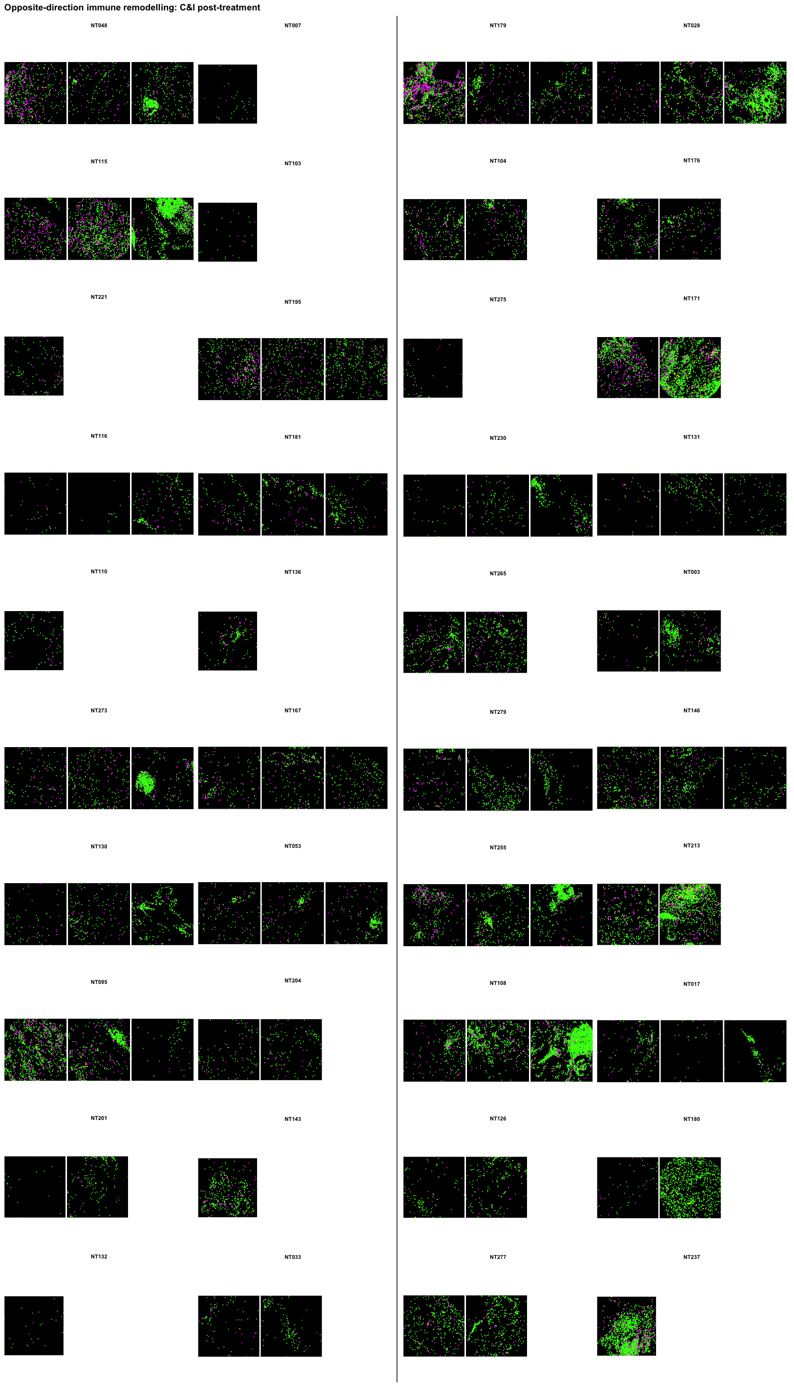
Figure 5 shows the two chemoimmunotherapy post-treatment phenotypes from the findings above on the tissue, for 20 representative patients per response group (those closest to each group’s median post-treatment M2 proportion). All post-treatment regions of interest are shown for each patient (pathological complete response on the left, residual disease on the right; magenta = M2 macrophages, yellow = PD-L1-positive antigen-presenting cells, green = other immune cells).

Spatial maps of post-treatment cores
cores <- read_csv(file.path(post_dir, "spatial-cores.csv"), show_col_types = FALSE)
cells <- read_fst(file.path(post_dir, "spatial-cells.fst")) |> as_tibble()

mk_roi <- function(image_id) {
  if (is.na(image_id)) return(patchwork::plot_spacer())
  mk_spatial_tile(filter(cells, ImageID == image_id))
}

mk_row <- function(pid) {
  image_ids <- cores |>
    filter(PatientID == pid) |>
    arrange(roi) |>
    pull(ImageID)
  ids <- c(image_ids, rep(NA_character_, 3L - length(image_ids)))
  patchwork::wrap_plots(
    ggplot() +
      annotate("text", x = 0.5, y = 0.5, label = pid, fontface = "bold", size = 2.8) +
      scale_x_continuous(limits = c(0, 1), expand = c(0, 0)) +
      scale_y_continuous(limits = c(0, 1), expand = c(0, 0)) +
      theme_void(),
    patchwork::wrap_plots(lapply(ids, mk_roi), nrow = 1, ncol = 3),
    ncol = 1,
    heights = c(0.16, 1)
  )
}

mk_block <- function(response, title) {
  one_column <- function(which_side) {
    pids <- cores |>
      filter(pCR == response, side == which_side) |>
      arrange(patient) |>
      distinct(PatientID) |>
      pull(PatientID)
    patchwork::wrap_plots(lapply(pids, mk_row), ncol = 1)
  }
  patchwork::wrap_plots(one_column("left"), one_column("right"), ncol = 2) +
    patchwork::plot_annotation(
      title = title,
      theme = theme(
        plot.title = element_text(face = "bold", size = 12, hjust = 0.5, margin = margin(b = 4))
      )
    )
}

divider <- ggplot() +
  annotate("segment", x = 0.5, xend = 0.5, y = 0, yend = 1, linewidth = 0.7, colour = "grey35") +
  scale_x_continuous(limits = c(0, 1), expand = c(0, 0)) +
  scale_y_continuous(limits = c(0, 1), expand = c(0, 0)) +
  theme_void()

patchwork::wrap_plots(
  mk_block("pCR", "pCR (responder)"),
  divider,
  mk_block("RD", "RD (non-responder)"),
  ncol = 3,
  widths = c(1, 0.03, 1)
) +
  patchwork::plot_annotation(
    title = "Opposite-direction immune remodelling: C&I post-treatment",
    theme = theme(plot.title = element_text(face = "bold", size = 13))
  )
Spatial maps of representative post-treatment tumours in the chemoimmunotherapy arm. M2 macrophages are bright magenta, PD-L1-positive antigen-presenting cells are bright yellow, and other immune cells are bright green, with pathological complete response on the left and residual disease on the right.
Figure 6

Post-treatment M2 levels alone look similar between the two groups (21.9% in pCR vs 18.2% in RD among the displayed patients; 22.8% vs 20.9% across the cohort). That is why the visit-only contrast is a poor summary, and why the difference-in-differences contrast was used.


Show code
sessionInfo()
R version 4.6.1 (2026-06-24)
Platform: aarch64-apple-darwin23
Running under: macOS Tahoe 26.6.2

Matrix products: default
BLAS:   /Library/Frameworks/R.framework/Versions/4.6/Resources/lib/libRblas.0.dylib 
LAPACK: /Library/Frameworks/R.framework/Versions/4.6/Resources/lib/libRlapack.dylib;  LAPACK version 3.12.1

locale:
[1] C.UTF-8/C.UTF-8/C.UTF-8/C/C.UTF-8/C.UTF-8

time zone: Australia/Sydney
tzcode source: internal

attached base packages:
[1] stats     graphics  grDevices utils     datasets  methods   base     

other attached packages:
[1] fstcore_0.10.0  tidyr_1.3.2     dplyr_1.2.1     readr_2.2.0    
[5] patchwork_1.3.2 here_1.0.2      fst_0.9.8       ggplot2_4.0.3  

loaded via a namespace (and not attached):
 [1] tidyselect_1.2.1            farver_2.1.2               
 [3] S7_0.2.2                    fastmap_1.2.0              
 [5] SingleCellExperiment_1.34.0 tensorA_0.36.2.1           
 [7] digest_0.6.39               lifecycle_1.0.5            
 [9] sccomp_2.5.2                processx_3.9.0             
[11] magrittr_2.0.5              posterior_1.7.0            
[13] compiler_4.6.1              sass_0.4.10                
[15] rlang_1.3.0                 tools_4.6.1                
[17] gt_1.4.0                    utf8_1.2.6                 
[19] yaml_2.3.12                 data.table_1.18.4          
[21] knitr_1.51                  S4Arrays_1.12.0            
[23] labeling_0.4.3              htmlwidgets_1.6.4          
[25] bit_4.6.0                   DelayedArray_0.38.2        
[27] xml2_1.6.0                  RColorBrewer_1.1-3         
[29] cmdstanr_0.9.0              abind_1.4-8                
[31] withr_3.0.3                 purrr_1.2.2                
[33] BiocGenerics_0.58.1         grid_4.6.1                 
[35] stats4_4.6.1                scales_1.4.0               
[37] SummarizedExperiment_1.42.0 cli_3.6.6                  
[39] rmarkdown_2.31              crayon_1.5.3               
[41] generics_0.1.4              otel_0.2.0                 
[43] tzdb_0.5.0                  stringr_1.6.0              
[45] parallel_4.6.1              XVector_0.52.0             
[47] matrixStats_1.5.0           vctrs_0.7.3                
[49] Matrix_1.7-5                jsonlite_2.0.0             
[51] callr_3.8.0                 IRanges_2.46.0             
[53] hms_1.1.4                   S4Vectors_0.50.1           
[55] bit64_4.8.2                 ggrepel_0.9.8              
[57] glue_1.8.1                  ggside_0.4.1               
[59] distributional_0.8.1        stringi_1.8.7              
[61] gtable_0.3.6                GenomicRanges_1.64.0       
[63] instantiate_0.2.3           tibble_3.3.1               
[65] pillar_1.11.1               htmltools_0.5.9            
[67] Seqinfo_1.2.0               R6_2.6.1                   
[69] rprojroot_2.1.1             vroom_1.7.1                
[71] evaluate_1.0.5              Biobase_2.72.0             
[73] lattice_0.22-9              backports_1.5.1            
[75] Rcpp_1.1.2                  SparseArray_1.12.2         
[77] checkmate_2.3.4             xfun_0.60                  
[79] MatrixGenerics_1.24.0       fs_2.1.0                   
[81] forcats_1.0.1               pkgconfig_2.0.3            

© 2025 tidyomics. Content is published under Creative Commons CC-BY-4.0 License for the text and BSD 3-Clause License for any code. | R-Bloggers